Diagnostic Imaging (Radiology)
The Radiology Department at Hazel Hawkins Hospital provides patients access to a full range of radiological diagnostic services, performed by our highly skilled staff using state-of-the-art equipment. The staff providing patient care is specially trained and has licensure and/or certification in their area of specialty, to ensure that the highest standard of care is afforded our patients. Our Radiologists are Board certified by the American College of Radiology.
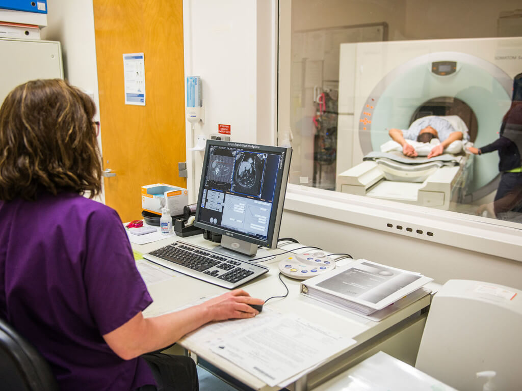
CT Multidetector Scanner
CT examinations, sometimes referred to as “CAT scans,” use a series of x-ray images – or slices – that are fed into a computer to produce detailed three-dimensional images to aid physicians in the diagnosis and treatment of disease and injury. Our CT scanner, which obtains 64 slices per rotation, significantly reduces scan times while improving patient comfort and reducing the radiation dose. Even more importantly, the multi-detector scanner allows staff to perform non-invasive examinations of virtually every system in the body, quickly and painlessly, with a level of digital detail that is no less than amazing. The scanner is utilized to provide inpatient, outpatient, and emergency diagnostic CT scans. In addition, it performs CT angiography and has cardiac imaging capabilities, promoting the ability to detect heart disease in its earliest stage.
MRI: (Magnetic Resonance Imaging)
Magnetic resonance imaging (MRI) is an imagining technique that uses a magnetic field, radio waves and a computer to compose detailed cross-sectional images of human anatomy. MRI’s produce better quality images of soft-tissue compared to x-rays. MRI’s are most commonly used to image the brain, spine, thorax, vascular system and musculoskeletal system.
The advantages of Hazel Hawkins Hospital’s MRI:
- High field strength for better image quality
- Shorter procedure times
- Wider range of procedures
- Images can be stored on a CD for easy portability back to referring physician.
Help for claustrophobic patients:
- Shorter bore scanner
- Short Tube
- Entrance to magnet is flared
- Patients can make arrangements prior to scan to tour MRI room and discuss procedure with technologist.
- Patients can discuss options with referring physician for mild sedative.
- For certain procedures, headphones with music are available.
- Use of prism glasses to reduce feeling of claustrophobia.
Bone Densitometry
Bone density scanning, also called dual-energy x-ray absorptiometry (DXA or DEXA) or bone densitometry, is an enhanced form of x-ray technology that is used to measure bone mineral loss. DEXA is today’s established standard for measuring bone mineral density (BMD). DEXA is most often performed on the lower spine, hips, or upper extremity.
Mammography
Hazel Hawkins Hospital is accredited by the American College of Radiology, and offers complete digital mammography services, breast ultrasound, and breast biopsies.
Ultrasound
Ultrasound, also known as sonography, is imaging that uses sound waves to produce images of organs, vessels and tissues in the body. A small hand-held device called a transducer is placed in contact with the patient’s skin at the area to be studied. The transducer emits high frequency sound waves that humans cannot hear. These sound waves pass through the body and send back echoes as they bounce off organs, vessels and tissues. With the help of computers, the echoes are then converted to form an image.
Ultrasound is safe to use for fetal imaging because it uses sound waves instead of radiation. Having an ultrasound when you are pregnant helps to interpret the fetal position, delivery date, gender and multiple babies. Our state-of-the-art equipment also allows us to capture 3D and 4D images of a fetus.
Ultrasounds are being used throughout medicine to help detect breast cysts and gallstones. They are also used to examine the liver, kidneys, pancreas, spleen, colon, and urinary bladder for tumors, inflammation, stones or cysts.
Echocardiography
Echocardiography is a procedure that uses sound waves to create a digital image of the heart. The exam is interpreted by a cardiologist. In addition to routine echocardiography, Hazel Hawkins Memorial Hospital also offers Stress Echocardiography and Dobutamine Stress Echocardiography capabilities.